Clinical Decision Support for Oncology

PredictRx™ provides phenotypic drug sensitivity testing on patient-derived cancer cells to support treatment selection in challenging clinical scenarios.

Performance Metrics

Positive predictive value

90%

Negative predictive value

100%

Drugs Tested

~60

Turnaround time

4 to 5 weeks

Clinical Evidence

PredictRx™ is based on technology originally developed by Imagen Therapeutics and validated through multiple clinical studies. The platform was refined over nearly a decade of research collaboration with leading UK cancer centres.

Early assay development was in ovarian cancer patients via the acquisition of ascites fluid drains. Five point dose response curves were converted to cumulative normalised cell death so that response profiles could be modelled by linear regression. This allowed us to develop a powerful AI discriminator model that can rank order any in vitro assay response against where that response lies in relation to all the other historical assay responses we currently hold in our database. This approach powerfully addresses the challenge of controlling for the fact that cell death is not being measured in the patient directly.
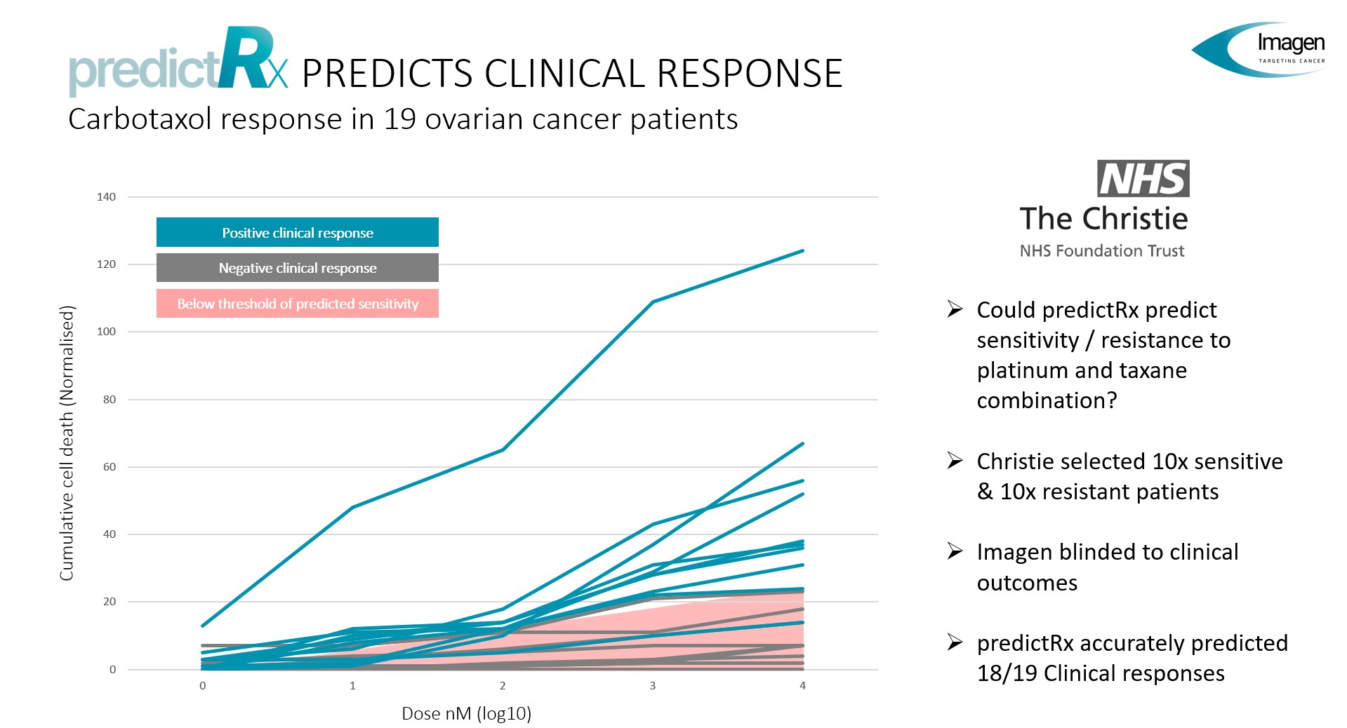
Additionally, before Imagen Therapeutics ceased trading in 2023, we had convincing data from 39 new patients where we had correctly predicted all 21 true negative clinical responses and all 16 positive responses. Out of the 39 patients, the assay only incorrectly assigned two patients with a positive response where the overall clinical outcome was judged to be negative. The table below summaries the 39 assays that were matched to clinical response.

Cancer Type Primary/Metastatic Correct Predictions Incorrect Predictions Drugs Red = Assay Non Response Green = Assay Response
Ovarian Metastatic 4 0 Gemcitabine Cisplatin Cisplatin/Paclitaxel Cisplatin/Etoposide
Ovarian Metastatic 2 1 Mitomycin C Paclitaxel Cisplatin
Ovarian (Ascites) Primary 18 1 Carbotaxel (10) Carbotaxel (9)
Fallopian Tube Carcinoma Primary 2 0 Cisplatin Cisplatin/Gemcitabine
Colorectal Metastatic 2 0 Irinotecan Oxaliplatin
CUP Metastatic 1 0 Cisplatin
Peritoneal Primary 1 0 Cisplatin/Paclitaxel
Ewings Sarcoma Primary 1 0 VIDE (Vincristine Ifosamide Doxorubicin Etoposide)
Kidney Metastatic 1 0 Sunitinib
Pancreatic Primary 1 0 Oxaliplatin
Breast Primary 1 0 Paclitaxel
Breast Metastatic 1 0 Lapatinib
Gastric Primary 1 0 Paclitaxel
Hepatocellular Carcinoma Metastatic 1 0 Sorafenib
Totals 39 37 2

Increasing Literature Support

Genotyping and proteomic analysis have been the dominate paradigms for personalising chemotherapy. However, although cancer is a genomic disease, little progress has been made using genotyping or proteomics to truly personalise chemotherapy. This is because there is an important difference between true personalised chemotherapy and stratified medicine. The former attempts to find the best available therapy from all the possible available chemotherapies to treat an individual patient. The latter tests whether a particular patient will respond to a particular chemotherapy regime. Genetic and proteomic testing are suited for stratified medicine, especially when the chemotherapy in question is a modern signal transduction targeting agent. 

For example, EGFR and BRAF inhibitors are known to be very effective in patients who carry certain critical signal transduction mutations. Yet while these tests are valuable in relation to specific drug treatments, we are yet to be able to analyse a complex cancer genetic or proteomic signature, and from that signature determine which chemotherapies will be beneficial to the patient.

Yet up until relatively recently, there was a general belief that attempting to personalise chemotherapy using phenotypic drug screening had been explored in the 1980s and 90s and the results were disappointing. Summarising the literature around these early studies it is possible to draw several important lessons.

  1. Early studies did not directly measure either cell death or cell number. Instead, the two most common approaches assessed ATP levels within plate wells or measured the conversion of the yellow MTT tetrazolium salt into purple formazan crystals. Both methods are influenced by cell number and metabolic activity, meaning that neither provides a direct measure of cell death or absolute cell count.
  2. A major review of this early work by Schrag (2004) found that, in many studies, patient response rates were significantly higher in assay-directed treatment groups than in groups receiving treatment based on physician choice alone. However, the authors’ principal criticism of this body of work was the lack of properly controlled randomised clinical trials. While this criticism has merit, it should also be recognised that demonstrating the value of a diagnostic assay does not fit neatly within the framework of a classical clinical trial, which is typically designed to evaluate whether a single new drug can treat a specific medical condition. For example, a trial that randomly assigns patients to either an assay-directed treatment group or a physician-choice group faces a major confounding factor: the intrinsic resistance of the tumour to treatment. The likelihood that either assay-guided selection or expert clinical judgement will identify an effective therapy is inversely related to the tumour’s overall resistance to chemotherapy.
  3. Paradoxically, personalised treatment strategies are most desirable for cancers with poor prognoses. Yet a poor prognosis often indicates that standard therapies for that cancer are already failing. In highly resistant cancers, where early attempts at phenotypic screening for treatment personalisation were often focused, statistical power was limited because chemosensitivity assays were effectively being asked to identify the best option among treatments that all had little or no therapeutic benefit.
  4. This consideration is particularly important because, since the 1990s, the number of available chemotherapies has increased substantially with the development of agents targeting specific signalling pathways. Moreover, because these modern therapies can be highly effective in small subpopulations of patients, assays capable of directly measuring whether a given treatment induces cancer cell death have become more important than ever.

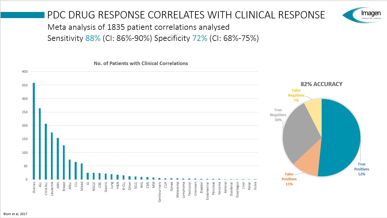
With these important background considerations, a meta analysis of 1835 patients correlations was published in 2017 by Blom et al demonstrating that despite the limitations imposed by the lack of viable treatment options, chemosenitivity assays still performed extremely well.
Recent Publications

Since the Blom publication in 2017, there have been an increasing number of publications (several in 2026) which indicate that chemosensitivity assays can play a vital role in optimising modern chemotherapy treatment. There is still a lot of discussion in the field over the the utility of 3D versus 2D cell culture. Imagen Therapeutics was able to work with both tissue configurations. Practically speaking, freshly prepared cancer biopsies will sometimes naturally clump and begin to grow in a more 3D-like organoid type configuration. When this occurred, we were able to process these structures using different High Content Analysis algorithms and also adopted a different cell death normalisation strategy to create a percentage cell death for the analysed spheroids.  The papers below give a flavour of the current status and progress of using chemosensitivity assay to personalise chemotherapy treatment (with links to pdf files when publicly available).

  1. Blom, K., Nygren, P., Larsson, R., & Andersson, C. R. (2017). Predictive Value of Ex Vivo Chemosensitivity Assays for Individualized Cancer Chemotherapy: A Meta-Analysis. SLAS Technology, 22(3), 306–314.
  2. J, van R. A. W., J, van de H., & Voest, E. E. (2023). Functional precision oncology using patient-derived assays: bridging genotype and phenotype. Nat Rev Clin Oncol, 20(5), 305–317.
  3. Letai, A., Bhola, P., & Welm, A. L. (2022). Functional precision oncology: Testing tumors with drugs to identify vulnerabilities and novel combinations. Cancer Cell, 40(1), 26–35.
  4. Loboda, A., Paluncic, J., Freitas, M., Weidmann, M., Herter, S., Zeiser, C., Schulz, J. P., Jády, A., Blattner-Johnson, M., Autry, R. J., Frese, K., Lupar, D., Schneider, M., Imle, R., Banito, A., Glimm, H., Witt, O., Peterziel, H., Kelm, J. M., … Oehme, I. (2026). Integration and validation of complementary ex vivo assays for functional precision oncology. NPJ Precis Oncol, 10(1), 230.This heroic study compares short-term ATP assays with more sophisticated long term organoid tissue culture and finds a high concordence between the two methods, especially in relation to cancers that possess defined oncogenic drivers.
  5. Ma, J., Liu, X., Xu, H., & Jiang, C. (2026). Patient-derived organoids in gastric cancer: bridging the tumor microenvironment to functional precision oncology. Front Bioeng Biotechnol, 14, 1790902.
  6. Mo, S., Luo, W., Yang, J., He, X., Ma, M., Cai, X., Li, Y., Hu, X., Mu, P., Cai, S., Ma, X., Liu, F., He, X., Zou, H., & Peng, J. (2026). Functional precision oncology platform of BRAF(V600E)-mutated colorectal cancer organoids predicts therapy response and reveals RNF43-mediated immunogenicity. Drug Resist Updat, 87, 101414.
  7. Panovska, D., & F, D. S. (2022). Functional Precision Oncology: The Next Frontier to Improve Glioblastoma Outcome? Int J Mol Sci, 23(15), 8637.
  8. Schrag, D., Garewal, H. S., Burstein, H. J., Samson, D. J., Van Hoff, D. D., & Somerfield, M. R. (2004). American Society of Clinical Oncology Technology Assessment: Chemotherapy Sensitivity and Resistance Assays. J.Clin.Oncol. 22, 3631-3638.This last reference was cited in an earlier discussion panel and is so included here for the reader's convenience.

Methodology

PredictRx™ employs a proprietary phenotypic drug sensitivity assay using low-passage patient-derived cells to maintain clinically relevant characteristics.

Technical Overview

  1. Sample Reception: Fresh tumour tissue is received and processed within 1-2 days of collection using a proprietary culture system.
  2. Cell Isolation and Culture: Tumour is enzymatically dissociated into single cells and cultured in selective media designed to preferentially support cancer cell growth while minimising normal cell contamination. Culture period: 3-4 weeks to achieve sufficient cell numbers (~2 million cells).
  3. Drug Screening: Cells are exposed to approximately 60 oncology drugs at 4 different concentrations each, generating dose-response curves for each compound. Assay duration: 72 hours.
  4. Analysis: Cell death is quantified using high-content imaging with nuclear staining. A computerised microscope scans all wells and measures relative cell death for each treatment.
  5. Computational Analysis: Results are analysed using proprietary machine learning algorithms and compared against a database of >150,000 drug-response data points from previous patients.
  6. Report Generation: Treatments are ranked and categorised based on relative efficacy. A clinical report is generated highlighting predicted effective and ineffective options.

Quality Control

Each assay includes positive and negative controls. Cell viability and purity are assessed before screening. Only samples meeting quality thresholds proceed to testing.

Final Imagen Therapeutics Drug Panel (2023)

PredictRx™ tested approximately 60 oncology agents across multiple drug classes. This was the final panel being used in 2023 and was regularly updated to reflect clinical practice.

Chemotherapies

  • 5-Fluorouracil
  • Bleomycin
  • Carmustine
  • Cisplatin
  • Cyclophosphamide
  • Dacarbazine
  • Docetaxel
  • Doxorubicin
  • Eribulin
  • Etoposide
  • Gemcitabine
  • Irinotecan
  • Lomustine
  • Melphalan
  • Methotrexate
  • Mitomycin C
  • Mitoxantrone
  • Oxaliplatin
  • Paclitaxel
  • Temozolomide
  • Teniposide
  • Topotecan
  • Vincristine
  • Vinorelbine

Targeted Therapies

  • Afatinib
  • Axitinib
  • Bortezomib
  • Bosutinib
  • Cabozantinib
  • Carfilzomib
  • Cediranib
  • Cladribine
  • Clofarabine
  • Crizotinib
  • Dabrafenib
  • Dasatinib
  • Epothilone B
  • Erlotinib
  • Everolimus
  • Ganetespib
  • Gefitinib
  • Herceptin
  • Imatinib
  • Lapatinib
  • Nilotinib
  • Olaparib
  • Osimertinib
  • Palbociclib
  • Pazopanib
  • Ponatinib
  • Rapamycin
  • Regorafenib
  • Ribociclib
  • Ruxolitinib
  • Sorafenib
  • Sunitinib
  • Trametinib
  • Vandetanib
  • Vemurafenib
  • Vismodegib
  • Vorinostat

Current Limitations

PredictRx™ does not currently test the following drug classes:

  • Immunotherapies: Checkpoint inhibitors (anti-PD-1, anti-PD-L1, anti-CTLA-4) and other immunomodulatory agents
  • Prodrugs: Agents requiring hepatic metabolism (e.g., capecitabine/Xeloda)
  • Anti-angiogenic monoclonal antibodies: Such as bevacizumab (Avastin)
  • CAR-T and other cell therapies: Require immune system context not present in vitro

Note: The assay tests direct cytotoxic effects on cancer cells and cannot model immune-mediated mechanisms or drug metabolism that occurs in vivo. We are actively developing capabilities to expand our testing into immunotherapy and CAR-T cell therapy areas. If you have specific therapeutic options you'd like us to evaluate, please contact us to discuss potential collaboration.

Sample Requirements & Logistics

Acceptable Sample Types

  • Solid Tissue: Minimum 1cm³ fresh tumour (preferred)
  • Core Biopsies: 4x 10mm cores (18G needle)
  • Ascites: 250ml minimum volume
  • Pleural Effusion: 250ml minimum volume

Post-treatment samples preferred for treatment-resistant disease. Treatment-naive samples acceptable for rare/undefined cancers.

Sample Handling

  • Fresh tissue required (no formalin fixation)
  • Store in provided NanoCool preservation kit
  • Ship via medical courier within 92 hours of collection
  • Maintain at room temperature (do not freeze)
  • Include completed clinical information form

Success Rates by Sample Type

  • Fresh surgical specimens: Highest success rate
  • Core biopsies: Variable (tissue type dependent)
  • Effusions: Good success for carcinomatosis
  • Post-radiation tissue: Lower viability

Not all samples yield viable cells. Success varies by tumour type, treatment history, and sample quality.

Turnaround Time

  • Sample to Lab: 1-2 days (courier)
  • Cell Culture: 3-4 weeks
  • Drug Screening: 3 days
  • Analysis & Report: 1-2 days
  • Total: Final report delivered within 4-5 weeks from sample receipt

Optimal Clinical Scenarios

PredictRx™ provides greatest clinical utility in specific situations where treatment decisions are challenging.

Rare Tumours

Undefined Standard of Care

When facing cancers with limited clinical trial data or established treatment algorithms, PredictRx™ can help identify potentially effective options from the broader oncology pharmacopeia.

Example: Rare sarcoma subtypes, unusual presentations of common cancers

Aggressive Disease

Treatment Optimization

For rapidly progressive disease where selecting the most effective treatment quickly is critical, PredictRx™ can help prioritise options within 3-4 weeks.

Example: High-grade neuroendocrine tumours, rapidly progressive metastatic disease

Refractory Disease

Treatment-Resistant Cancer

When cancer has progressed through standard therapies, PredictRx™ tests using treatment-resistant cells to identify remaining effective options.

Example: Platinum-resistant ovarian cancer, multiply-relapsed leukaemia

Several case studies have shown PredictRx™ correctly identifying EGFR inhibitor sensitivity in NSCLC later confirmed to have EGFR mutations, as well as predicting lack of EGFR sensitivity in EGFR wild-type cases — demonstrating potential utility as a functional biomarker.

Scroll to Top